


The Thoracic Spine: The Most Undertreated Region in the Body — and Why It’s Driving More Than Just Mid-Back Pain
If you have mid-back stiffness, you probably also have one or more of these: neck pain, forward head posture, shoulder tightness, low back ache, poor shoulder mobility, or headaches. You may have been treated for some of them. You may have been told they’re separate problems.
They’re not separate problems. They’re the same problem expressing itself in multiple places — and the thoracic spine is usually the origin.
The thoracic spine — the twelve vertebrae from the base of your neck to the bottom of your rib cage — is the most mechanically influential and most clinically misunderstood region of the spine. It attaches to every rib. It sits between the cervical spine and the lumbar spine. It houses the joints that allow your trunk to rotate. And critically, it is the home of the rib cage — the structure your diaphragm attaches to, which means it controls how you breathe.
When the thoracic spine isn’t moving correctly, everything connected to it compensates. That’s not a metaphor — it’s mechanics. Here’s how it works.
What the thoracic spine is actually for
The thoracic spine is twelve vertebrae, T1 through T12, each articulating with one or two pairs of ribs through the costovertebral and costotransverse joints. This rib attachment is what makes the thoracic spine mechanically unique — and what makes it both the most stable and the most complex region of the spine.
That stability is often described as the thoracic spine’s defining characteristic. You’ll read in many sources that the thoracic spine is “designed for stability” while the lumbar spine is for power and the cervical spine is for mobility. This is an oversimplification that has misled a lot of treatment decisions.
The thoracic spine is designed for rotation. It is the primary contributor to trunk rotation in activities like throwing, swinging, reaching, and changing direction. It is also designed for a specific degree of flexion — the normal thoracic kyphosis — that positions the rib cage correctly for breathing, shoulders correctly for overhead function, and the head correctly over the center of mass.
What it is not designed for is static extension, which is exactly where most people end up spending most of their time.
The shape problem — and why “sit up straight” makes it worse
Here is the central clinical insight that most patients have never been told, and that most treatment approaches don’t account for:
A normal thoracic kyphosis is not a postural defect. It is an anatomical requirement.
The thoracic spine is supposed to curve forward. This curvature positions the rib cage in its optimal three-dimensional shape — what respiratory physiologists and the Postural Restoration Institute describe as the zone of apposition — the position in which the diaphragm can function as a true piston rather than a flattened sheet. It allows the shoulder blades to sit flat against the rib cage. It positions the cervical spine so the head can be balanced over the center of mass without muscular effort. And it maintains appropriate lumbar lordosis below.
When patients are told to “sit up straight” or “pull your shoulders back,” the typical response is to extend the mid-thoracic spine — particularly at the T3-T5 region. This is exactly the wrong movement in exactly the wrong place. This region is frequently already locked in extension. Driving it further into extension doesn’t improve posture. It:
- Elevates and flares the rib cage, compressing the diaphragm
- Increases lumbar extension below (the pelvis tips forward, the lower back arches)
- Forces the lower cervical spine into flexion and the upper cervical spine into extension to keep the eyes level with the horizon
The result is the classic forward head posture — not from slouching, but from the attempt to stand up straight. The head isn’t forward because the person has poor habits. The head is forward because the mid-thoracic spine has locked in extension, the compensation patterns have propagated up and down the chain, and the entire system has reorganized around a fixed structural fault at T3-T5.
Flat back syndrome — the clinical diagnosis for loss of normal thoracic kyphosis — is a well-recognized pathological condition. The research on it is consistent: insufficient thoracic kyphosis produces worse clinical outcomes than normal or even moderately increased kyphosis. The spine needs its curves. Trying to eliminate them is not a therapeutic goal — it’s a misunderstanding of spinal anatomy.
Breathing: why it matters more than most providers realize
We breathe approximately 20,000 times per day. The mechanical demands of that are enormous, and the position of the rib cage determines whether breathing is effortful or efficient.
The diaphragm is the primary muscle of respiration. It attaches to the lower thoracic spine (T12), the lower six ribs, and the xiphoid process of the sternum. In its optimal position, the diaphragm forms a dome shape — highest in the center, descending at the periphery. On inhalation, it contracts and descends, increasing the volume of the thoracic cavity and drawing air in. This is diaphragmatic breathing. It’s efficient, low-effort, and mechanically sound.
The rib cage position determines whether the diaphragm can achieve and maintain this dome shape. When the rib cage is elevated and flared — which is what happens when the thoracic spine is locked in extension — the diaphragm is pulled into a flattened position. A flat diaphragm cannot descend efficiently. Inhalation becomes effortful. The accessory breathing muscles — the scalenes, the sternocleidomastoid, the upper trapezius — are recruited to help lift the rib cage on every breath.
Those muscles run from the upper ribs to the cervical spine. When they’re recruited as accessory breathing muscles 20,000 times a day, they become chronically overactive. Chronically overactive scalenes and upper trapezius are a primary driver of upper cross syndrome — the pattern of tight upper traps and pec minor combined with weak deep cervical flexors and lower trapezius that produces neck pain, shoulder blade winging, and cervicogenic headache.
Rib flare is the visible consequence of this pattern. When the lumbar spine hyperextends and the mid-thoracic spine locks in extension, the lower ribs are driven upward and outward — the rib cage can no longer close down in front. This is not a structural deformity. It’s a positional consequence of the spinal pattern above and below it. And it has an immediate effect on breathing: with the lower rib cage unable to descend and expand, diaphragmatic breathing is effectively turned off. The body compensates with cervical paradoxical breathing — the chest rises, the accessory muscles in the neck and upper shoulders lift the rib cage on every inhalation, and the diaphragm becomes a passenger rather than a driver. The result is the chronically tight neck and upper trapezius that patients describe as “where they carry their stress” — which is accurate, but incomplete. They’re carrying the mechanical cost of a breathing pattern that has no other option given the rib cage position they’re in.
Due to the inherent asymmetry of the human body — the liver sitting on the right, the left diaphragm working at a mechanical disadvantage — this pattern is almost never bilateral. Left rib flare is more common than right. The body rotates slightly to recruit the right side more effectively, which is why thoracic and pelvic asymmetries follow a consistent pattern across the population rather than being random. A patient presenting with left-sided rib flare, left-sided neck tension, and right-sided low back pain is not showing you three separate problems. They’re showing you one pattern with three expressions.
This is why treating upper cross syndrome without addressing the rib cage position and breathing pattern underneath it produces temporary results. The breathing pattern that’s driving the muscle overactivation is still there. The muscles return to their overactive state because they’re still being recruited as accessory breathing muscles thousands of times a day.
The rib cage is also connected to the pelvis through the abdominal canister — the diaphragm above, the pelvic floor below, and the abdominal wall surrounding. When the rib cage position is wrong, the abdominal canister cannot generate appropriate intra-abdominal pressure. This affects lumbar stability, loading patterns through the lumbar discs, and the activation patterns of the glutes and hip stabilizers. A mid-thoracic restriction doesn’t just cause mid-back pain. Through the rib cage and breathing mechanics, it contributes to low back pain, hip dysfunction, and pelvic floor issues — all from the same root.
The compensation cascade — what happens above and below
When a section of the thoracic spine loses rotation and flexion mobility — particularly in the T3-T8 region where restriction is most common — the segments above and below are forced to compensate.
Above: the cervical spine takes the load
The cervical spine is built for mobility. It has six degrees of freedom at each level and is capable of substantial range of motion in all directions. When thoracic rotation is restricted, cervical rotation is recruited to compensate — the neck rotates further than it should to allow the person to look over their shoulder, reach across their body, or simply function in a three-dimensional world.
Over time, this overloads the lower cervical segments — typically C4-C7, which are already the highest-load levels in the neck. The result is:
- Neck pain and stiffness that doesn’t fully resolve with cervical treatment alone
- Cervicogenic headache — headaches originating from the upper cervical spine, driven by the chronic overload of that region
- Forward head posture — the head migrates anterior to the center of mass as the compensation pattern becomes fixed
- Upper trapezius and levator scapulae tension — chronic protective guarding around the overloaded cervical segments
The clinical implication: a patient presenting with neck pain who has thoracic restriction will not achieve lasting improvement from cervical manipulation alone. The neck is the compensator. The thoracic spine is the cause.
Below: the lumbar spine loses its base
The lumbar spine requires a stable, mobile thoracic spine above it to function correctly. When thoracic rotation is restricted, lumbar rotation is recruited to compensate — particularly in activities involving trunk rotation. The lumbar spine is not designed for rotation. It has very limited rotational range of motion by design, and what little it has is not meant to carry the load that should be distributed through thoracic rotation.
The chronic demand for lumbar rotation compensation produces facet joint overload, disc loading in shear, and the muscular guarding patterns associated with low back pain.
Additionally, the mid-thoracic extension pattern described above drives anterior pelvic tilt below — the pelvis tips forward as the lumbar spine hyperextends to compensate, which shortens the hip flexors, inhibits the glutes, and loads the lumbar discs in extension. This is the same pattern that defines lower cross syndrome — and it is frequently driven, at least in part, from the thoracic spine above.
The shoulder: scapular positioning depends on rib cage shape
The scapula sits on the posterior rib cage and depends on the shape of that rib cage to position correctly. When the rib cage is elevated and flared — the consequence of thoracic extension and poor breathing mechanics — the scapula cannot sit flush against it. The result is scapular winging, altered scapulohumeral rhythm, and reduced subacromial space on shoulder elevation.
This is one of the most common underlying contributors to rotator cuff impingement and shoulder pain that isn’t resolved by rotator cuff strengthening exercises alone. If the rib cage the scapula is sitting on is the wrong shape, the scapula is in the wrong position regardless of how strong the rotator cuff is.
Rib subluxation — the thoracic spine’s most common acute presentation
The costovertebral and costotransverse joints — where the ribs attach to the thoracic spine — are the most frequently subluxated joints in the mid-back. When thoracic mobility is restricted, these joints bear the mechanical cost of every breath and every rotation.
Rib subluxation produces sharp, localized pain with breathing, twisting, or sneezing — often mistaken for cardiac, pulmonary, or gastrointestinal problems because the pain can radiate anteriorly toward the sternum. It responds quickly to specific chiropractic adjustment of the costovertebral joint, but recurs when the underlying thoracic restriction driving the abnormal rib mechanics is not addressed.
Recurrent rib subluxation is almost always a thoracic mobility problem, not just a rib problem.
What conventional treatment gets wrong
The standard approach to mid-back pain — in chiropractic, physical therapy, and online ergonomic advice — centers on two interventions:
Thoracic extension manipulation. The traditional chiropractic approach to the thoracic spine involves placing the patient prone or supine and applying a high-velocity thrust in the extension direction. This produces cavitation (the pop) at the targeted joints and often provides immediate local pain relief.
The problem is that it addresses the wrong direction of motion. Thoracic restriction is primarily a loss of rotation and flexion — not a loss of extension. The thoracic spine that is stuck in extension at T4 does not need more extension. It needs rotation and flexion restored at that segment and the segments around it. Repeatedly thrusting the thoracic spine into extension may provide temporary symptom relief while reinforcing the very mechanical pattern driving the problem.
This is not a condemnation of thoracic manipulation — it is a valuable tool when applied correctly, in the right direction, at the right level. It is a critique of the reflexive application of extension-based manipulation to a spine that is already in extension.
“Sit up straight” postural advice. As described above, the conventional advice to extend the thoracic spine and retract the scapulae to improve posture drives the mid-thoracic extension pattern further. Most ergonomic advice, most postural correction cues, and most “good posture” instruction available online operates on a model of the spine that does not account for the three-dimensional shape requirements of the thoracic cage or the role of breathing mechanics in maintaining spinal position.
The patient who has been doing postural exercises, trying to sit up straight, and getting regular thoracic adjustments without lasting improvement is not failing at their rehabilitation. They are being given an incomplete framework.
What the assessment should look for
A thorough evaluation of mid-back pain and thoracic dysfunction should include:
Rotational mobility assessment — not just gross trunk rotation, but segmental assessment of which thoracic levels are contributing to restriction and in which direction. This is different from a standard range of motion measurement.
Rib cage position and breathing mechanics — does the rib cage elevate and flare on inhalation rather than expanding three-dimensionally? Does the patient breathe primarily into the chest with accessory muscle recruitment rather than into the lower rib cage? These findings change the treatment approach.
Scapular position and kinematics — does the scapula sit flush against the rib cage? Is there asymmetry in resting position? Does it move correctly through shoulder elevation or does it wing and tip?
Cervical and lumbar compensation patterns — what has the thoracic restriction produced above and below? Is the neck pain or low back pain a compensator for the thoracic finding, or an independent problem?
The overall postural organization — where is the patient’s center of mass? What is the relationship between rib cage position, pelvic position, and head position? This requires looking at the whole system, not just the region that hurts.
How we treat it at Ashworth
The thoracic spine is one of the regions where the difference between a thorough clinical framework and a routine adjustment is most apparent in outcomes.
Our approach starts with the assessment above — identifying which levels are restricted, in which direction, and what the rib cage and breathing pattern are contributing to the picture. Treatment is then directed at restoring the specific mobility that’s missing:
- Chiropractic mobilization and manipulation specific to the restricted segment and direction — rotation and flexion restoration, not reflexive extension thrusting
- Rib joint mobilization — addressing the costovertebral and costotransverse joints that are restricted by the same pattern
- Soft tissue work — the thoracic paraspinals, rhomboids, serratus anterior, and intercostals all need attention when thoracic mobility is chronically restricted
- Breathing mechanics rehabilitation — restoring the diaphragmatic breathing pattern and rib cage position that the thoracic restriction has compromised. This is not just a nice addition to treatment — it is often the difference between improvement that holds and improvement that recurs
- Shockwave therapy (ESWT) — where thoracic restriction has produced persistent trigger points in the paraspinal musculature or intercostal region, shockwave therapy addresses the soft tissue component that manual therapy alone may not fully resolve
The Postural Restoration Institute (PRI) framework provides the clinical model that integrates all of these elements. PRI treats the thoracic spine as part of a three-dimensional system — accounting for the inherent asymmetry of human anatomy, the role of the rib cage in breathing and postural organization, and the compensation patterns that develop above and below a restricted thoracic spine. It is the missing piece that makes the difference between treatment that provides temporary relief and treatment that actually changes the underlying pattern.
Most patients with mid-back stiffness and thoracic restriction — even longstanding cases — respond well to a focused course of care. Our average across all musculoskeletal conditions is 6.3 visits to resolution. The thoracic spine, when properly assessed and treated, responds faster than most patients expect.
If you’re in the Des Moines or West Des Moines area and dealing with mid-back pain, thoracic stiffness, or any of the downstream patterns described on this page, reach us at (515) 225-4002 or schedule an appointment online. No referral needed.
Frequently asked questions
What causes mid-back pain and stiffness?
Mid-back pain most commonly comes from thoracic joint restriction — particularly loss of rotation and flexion mobility at specific segments. Sustained postures, rib joint dysfunction, and compensatory patterns from the neck or lower back are the most common drivers. In many cases the thoracic spine has become locked in extension at one area, forcing the segments above and below to compensate with excessive flexion — a pattern that produces pain both locally and at a distance.
Can mid-back stiffness cause neck pain?
Yes — and this is one of the most commonly missed connections in musculoskeletal care. When the mid-thoracic spine loses rotation mobility, the cervical spine compensates by moving more. Over time this overloads the lower cervical segments and produces neck pain, cervicogenic headache, and upper trapezius tension that won’t resolve with neck treatment alone. Treating the neck without addressing the thoracic restriction is treating the compensator, not the cause.
Why does standing up straight make my posture worse?
Because “standing up straight” typically means extending the mid-thoracic spine — which is usually already stuck in extension. The result is increased lumbar arching below and cervical flexion above, which pushes the head further forward. A normal thoracic kyphosis is not a postural flaw. Flat back syndrome — the loss of thoracic kyphosis — is an actual clinical diagnosis with worse outcomes than normal kyphosis.
How does the thoracic spine affect breathing?
The diaphragm attaches to the lower thoracic spine and lower ribs. When the thoracic spine is stuck in extension and the rib cage is elevated and flared, the diaphragm is compressed into a flat position and cannot descend properly on inhalation. Accessory breathing muscles in the neck and shoulders compensate — and when they’re recruited 20,000 times a day, they become chronically overactive. This drives neck tension, upper cross syndrome, and contributes to shoulder and low back pain through altered trunk mechanics.
Why doesn’t thoracic manipulation fix mid-back stiffness long-term?
Because most thoracic manipulation pushes the spine into extension — the direction it’s often already restricted in. It provides temporary relief but doesn’t restore the rotation and flexion mobility that’s missing, and doesn’t address the rib cage position or breathing pattern maintaining the problem. Long-term improvement requires direction-specific mobilization, rib mobility work, and correction of the breathing and postural patterns driving the dysfunction.
upuncture, and customized stretches and exercises for home care.
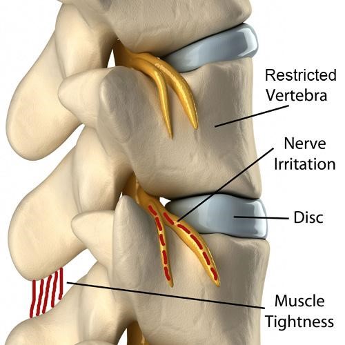
Image on this page provided by ChiroUp.
Your Path to Wellness Begins Here!
Our experts in chiropractic, physical therapy, acupuncture, and massage therapy work as a team to provide holistic care that meets your specific needs. Book your appointment today and experience coordinated care.
I really love it here! They are very friendly and I feel Dr. Wilson is giving me great treatment! I also don’t feel pressured into coming back too frequently. The adjustments are great! Great office!
Very professional and in depth consultation. These people seem to genuinely care about people’s health and progress.
Dr. Matt has done wonders for me and I recommend him highly – I can’t say enough good things about him. Dr. Matt and staff are good people and will take great care of you.
My family have used chiropractic and physical therapy services here and every one of us have had excellent outcomes. I wouldn’t go anywhere else these folks are as good as it gets.
Dr. Matt and the entire staff have been GREAT! I went in for some issues I was having with my neck and back and Dr. Matt also helped me with some other issues I was having with my calf and shoulders that have made a HUGE improvement in my overall health and comfort.I highly recommend using Dr. Matt!
Dr. Matthew squeezed me in on short notice and took very good care of me. He was very thorough and didn’t just adjust me and let me walk out. Amazing experience here and the receptionist is a sweetheart.