


Radicular Pain: What It Is, What Causes It, and How to Get Rid of It
Your doctor called it radicular pain. Maybe they said radiculopathy. Maybe they said you have a pinched nerve, and the imaging shows a herniated disc at L4-L5 or C6-C7. Or maybe you haven’t seen anyone yet — you just know that there’s pain shooting down your arm or leg and it doesn’t feel like anything you’ve had before.
Whatever brought you here: this is a well-understood condition with a clear mechanical explanation, and the majority of people get better without surgery. Here’s what’s actually going on.
What radicular pain actually is
Radicular pain is nerve pain that starts in the spine and travels along a nerve root into your arm or leg.
Your spinal cord exits the skull and runs down through your vertebrae. At each level, nerve roots branch off left and right and travel outward to supply sensation and motor control to specific parts of your body. When one of those nerve roots gets compressed or irritated — by a herniated disc, a bone spur, or a narrowed spinal canal — it generates pain, numbness, tingling, or weakness that follows the path of that nerve rather than staying localized at the spine.
This is what distinguishes radicular pain from ordinary low back pain or neck pain. Ordinary back pain stays where the problem is. Radicular pain travels — often a long way from where the actual compression is occurring.
The specific path it follows depends on which nerve root is affected. This is called a dermatomal distribution — each nerve root corresponds to a predictable region of skin and muscle. A compressed L4 nerve root produces a different pattern than a compressed L5 or S1. A compressed C6 nerve root travels differently than C7 or C8. This predictability is part of what makes radicular pain diagnosable and, importantly, treatable.
What it feels like
The sensation is usually distinct enough that patients recognize it as different from other pain they’ve experienced. Common descriptions:
- Shooting or electric pain that travels from the neck or lower back into the arm or leg — sometimes all the way to the hand or foot
- Burning along the path of the nerve
- Numbness or tingling — often in a specific region of the hand, forearm, thigh, calf, or foot
- Muscle weakness in the affected limb — difficulty gripping, foot drop, weakness pushing off when walking
- Pain that worsens with specific positions — sitting, bending forward, sneezing, coughing, or bearing down, depending on which nerve is involved
- Pain that improves with movement or with positions that open the neural foramina (the exit points where nerve roots leave the spine)
One thing that catches people off guard: the pain in the arm or leg can be significantly worse than any pain at the spine itself. A patient with L5-S1 radiculopathy may have minimal low back pain but excruciating leg pain. The spine is the source; the leg is where it expresses.
Cervical vs. lumbar radicular pain — the two versions
Lumbar radicular pain (into the leg) is the more common form. Sciatica — pain traveling from the low back through the buttock and into the leg — is the best-known version. The most commonly affected levels are L4-L5 and L5-S1, which supply the gluteal region, hip abductors, thigh, calf, and foot. Lumbar radiculopathy can produce pain, numbness, or weakness anywhere along that path.
Cervical radicular pain (into the arm) originates in the neck. The most commonly affected levels are C5-C6 and C6-C7, which produce pain, numbness, or tingling into the shoulder, arm, forearm, and hand. Patients sometimes describe it as pain down the arm combined with a specific patch of numbness — often in one or two fingers — that helps localize which nerve root is involved.
Both follow the same basic mechanism: nerve root compression or irritation. The structural cause and the treatment approach are similar; the location of symptoms is what differs.
What causes it
Herniated disc — the most common cause, particularly in adults under 50. Each spinal disc has a tough outer ring (annulus fibrosus) and a gel-like center (nucleus pulposus). When the outer ring develops a tear or weakens, the inner material can bulge or extrude outward and press against an adjacent nerve root. Herniated discs can occur from a single event — a heavy lift, a sudden twist — or develop gradually from repetitive loading.
Degenerative disc disease and bone spurs — more common in adults over 50. As discs lose height with age, the vertebrae move closer together and the body responds by forming bone spurs (osteophytes) at the margins of the vertebrae and facet joints. These can narrow the neural foramina and compress nerve roots. This is a gradual process, which is why cervical radiculopathy from degenerative changes tends to develop more slowly than disc herniation-related cases.
Spinal stenosis — narrowing of the spinal canal itself, usually from a combination of disc bulging, facet joint hypertrophy, and ligament thickening. Central stenosis can compress multiple nerve roots at once. Lateral recess stenosis narrows the exit points where individual nerve roots leave. Stenosis-related radicular pain often has a characteristic feature: it worsens with walking or standing (positions that extend the spine) and improves with sitting or bending forward (positions that open the canal).
Foraminal stenosis — narrowing specifically at the neural foramen, the opening where the nerve root exits the spine. This is one of the most common structural findings on MRI in patients with cervical and lumbar radiculopathy, and it’s one of the most responsive to conservative care because the foramen can be mechanically opened with specific positioning and joint mobilization.
Spondylolisthesis — when one vertebra slips forward relative to the one below it, the resulting instability and altered mechanics can compress nerve roots at that level.
What the imaging shows — and what it doesn’t tell you
Most patients with radicular pain get an MRI at some point, and many arrive with their results already in hand. A few things worth understanding about imaging findings:
The MRI tells you what’s there, not necessarily what’s causing your symptoms. Studies consistently show that herniated discs, degenerative disc disease, and even nerve root compression are common findings on MRI in people with no symptoms at all. The imaging finding has to match your clinical presentation — the dermatomal pattern, the neurological exam, the provocative testing — for it to be meaningful.
Most herniated discs reabsorb. There is strong evidence that a significant portion of disc herniations, including large ones, decrease in size over time as the immune system clears the extruded disc material. This is one of the reasons the majority of patients with lumbar disc herniation improve without surgery — the structural problem often resolves on its own, especially with appropriate conservative care.
Imaging doesn’t tell you how to treat it. The same L4-L5 herniation looks identical on MRI whether the patient has been symptomatic for two weeks or two years, whether the nerve root is highly irritated or mildly compressed, and whether the patient needs aggressive care or minimal intervention. The clinical picture — exam findings, symptom severity, functional limitations, and response to treatment — guides management far more than the image.
The difference between radicular pain, radiculopathy, and referred pain
These terms get used interchangeably but they’re not the same:
Radicular pain refers specifically to the pain itself — the shooting, traveling, dermatomal pattern that follows a nerve root.
Radiculopathy is a broader term that refers to dysfunction of a nerve root — which may include radicular pain, but also encompasses numbness, weakness, and reflex changes. A patient can have radiculopathy with significant weakness but minimal pain. Or significant pain but no measurable neurological deficit.
Referred pain is different from both. Referred pain (often called piriformis syndrome) is a non-dermatomal aching pattern generated by muscles, ligaments, and joints — not nerve roots. Trigger points in the gluteus medius can refer pain down the leg in a pattern that looks like sciatica. Facet joints can refer pain into the hip or thigh. The distinction matters because treatment is different. Referred pain that’s been diagnosed as radiculopathy is one of the more common misdiagnoses in musculoskeletal care.
A thorough clinical examination should be able to distinguish between them. The nerve tension tests (straight leg raise, slump test, upper limb tension tests), dermatomal sensory testing, reflex testing, and myotomal strength testing give enough information to differentiate true nerve root involvement from referred pain in most cases.
Conservative care — what works and why
The research on conservative care for radicular pain is clear: the majority of patients improve without surgery, and the prognosis for most forms of radiculopathy with appropriate conservative management is good.
Chiropractic care is among the most evidence-supported conservative treatments for lumbar and cervical radiculopathy. Spinal manipulation and joint mobilization address the mechanical component of nerve root compression — particularly foraminal stenosis and disc-related compression — by restoring normal joint mobility, reducing inflammatory pressure on the nerve root, and improving the mechanical environment around the affected segment. Cox Flexion-Distraction, a specific technique that decompresses the intervertebral disc and neural foramina under traction, has strong clinical evidence for lumbar disc herniation and radiculopathy. The neural foramina can be mechanically opened with the right technique — this is not just symptom management, it addresses a structural component of the problem.
Physical therapy contributes through targeted rehabilitative exercise that stabilizes the affected spinal segment, addresses muscular imbalances that may be contributing to aberrant loading, and restores functional movement patterns. For lumbar radiculopathy specifically, directional preference exercises (McKenzie method) — finding the movement direction that centralizes symptoms — are well-supported and often produce rapid improvement.
Acupuncture has an accumulating evidence base for radicular pain, primarily through modulation of inflammatory mediators around the nerve root and central sensitization pathways. For patients with significant inflammatory component to their radiculopathy, acupuncture can meaningfully reduce pain levels and is a useful adjunct to manual care.
Shockwave therapy (ESWT) has an emerging and clinically meaningful role in radicular pain, particularly in presentations where soft tissue is the primary driver rather than disc or bone. Two specific situations where it fits well:
- Piriformis syndrome — when the sciatic nerve is being compressed by the piriformis muscle rather than a spinal disc, the MRI often looks unremarkable and patients are frequently misdiagnosed with lumbar disc radiculopathy. The pain pattern can be nearly identical. Shockwave therapy directed at the piriformis muscle reduces muscular tension, breaks down fibrous adhesions around the sciatic nerve, and addresses trigger points that are mechanically compressing the nerve. Patients who’ve had lumbar-focused treatment without improvement are worth evaluating for this presentation specifically.
- Failed back surgery syndrome and post-surgical scar tissue — patients who’ve had lumbar surgery and continue to have radicular symptoms despite a structurally successful procedure are a particularly difficult population. Epidural fibrosis — scar tissue that forms around the nerve roots following surgery — is a common and underappreciated cause of persistent radicular pain post-operatively. Shockwave therapy can address the soft tissue component of this, reaching tissue that manual therapy cannot. It doesn’t undo the surgery, but it can meaningfully reduce the residual nerve irritation that persists when everything else looks structurally fine.
We use the Storz Duolith SD1 Ultra — one of two focused shockwave devices of this kind in Iowa and the only one in the Des Moines metro. The focused device matters here: it delivers energy precisely to the target tissue at depth, which is what piriformis work and post-surgical cases require. Radial devices used by most clinics cannot reach the depth or precision needed for these presentations.
Traction — either mechanical or manual — decompresses the neural foramina and reduces intradiscal pressure. It’s particularly useful in foraminal stenosis and acute disc herniation cases where the nerve root is under direct mechanical load.
The combination of approaches matters. Patients who receive manual care alongside rehabilitative exercise consistently show better outcomes than those receiving either alone.
When conservative care isn’t the right answer — and what signals to watch for
Most radicular pain cases belong in conservative care. But a few situations require immediate medical evaluation:
Post-surgical radicular pain deserves its own note. If you’ve had lumbar surgery and still have nerve pain down your leg, the answer isn’t necessarily more surgery. Epidural fibrosis, residual foraminal stenosis, and altered biomechanics from the surgical level are all treatable conservatively. We see this population regularly and it’s worth a thorough evaluation before assuming the only path forward is another procedure.
Cauda equina syndrome — if you have radicular symptoms combined with loss of bowel or bladder control, saddle anesthesia (numbness in the groin/inner thighs), or rapidly progressing weakness in both legs, this is a surgical emergency. Don’t wait for a chiropractic appointment. Go to an emergency room.
Progressive neurological deficit — weakness that is measurably worsening over days to weeks, rather than stable or improving, warrants urgent evaluation. A nerve root that’s being progressively damaged needs surgical decompression before permanent deficit occurs.
Failure of conservative care — if symptoms haven’t improved after 6–12 weeks of appropriate conservative management, surgical consultation is reasonable. We don’t hold patients in conservative care indefinitely when it’s not working.
For everything else — which is the vast majority of radicular pain presentations — conservative care is the evidence-based starting point, not the fallback after everything else fails.
How we approach this at Ashworth
Radicular pain is one of the conditions we see most often, and it’s one where getting the diagnosis right before starting treatment makes a significant difference in how quickly patients improve.
The evaluation includes nerve tension testing, dermatomal sensory and strength assessment, reflex testing, and clinical correlation with any available imaging. The goal is to confirm that we’re dealing with true radiculopathy rather than referred pain, identify which nerve root is involved and what’s driving the compression, and determine whether there are any red flags that would indicate this isn’t the right setting for care.
From there, the treatment approach depends on the structural diagnosis. Foraminal stenosis and disc herniation respond differently than central stenosis. Acute presentations are managed differently than chronic or recurrent ones. We match the technique to the finding rather than applying the same protocol to every back or neck complaint that comes through the door.
Most patients with radicular pain do not need a long treatment course. Our average across all musculoskeletal conditions is 6.3 visits to resolution. For acute disc herniation with radiculopathy, many patients see significant symptom centralization and improvement within the first 3-8 visits as the acute inflammatory response settles and the mechanical pressure on the nerve root is reduced.
If you’re in the Des Moines or West Des Moines area and are dealing with nerve pain down your arm or leg, reach us at (515) 225-4002 or schedule an appointment online. We’ll tell you honestly what we’re seeing on examination, what we think the timeline looks like, and whether we’re the right fit for what’s going on.
Frequently asked questions
What is radicular pain?
Radicular pain is nerve pain that originates in the spine and travels along a nerve root into your arm or leg. It’s caused by irritation or compression of a spinal nerve root — most commonly from a herniated disc, bone spur, or spinal stenosis. The pain follows a predictable dermatomal path based on which nerve is affected. Sciatica is the most familiar form, but radicular pain can occur in the neck and travel into the arm as well.
What does radicular pain feel like?
Most people describe shooting, electric, or burning pain that travels from the neck or back into the arm or leg, often following a specific path. Numbness and tingling in a particular region — one or two fingers, the outer calf, the bottom of the foot — are common and help identify which nerve root is involved. Muscle weakness can accompany severe cases. Symptoms often worsen with sitting, coughing, or sneezing and may improve with walking or specific positional changes.
Is radicular pain the same as sciatica?
Sciatica is a specific type of radicular pain involving the sciatic nerve, which originates from nerve roots L4 through S1 in the lumbar spine. Radicular pain is the broader term — sciatica is the most common lumbar version. Cervical radicular pain travels into the arm rather than the leg and involves different nerve roots, but the underlying mechanism is the same.
Can radicular pain go away without surgery?
Yes — the majority of radicular pain cases resolve with conservative care. Research consistently shows that most patients with herniated disc-related radiculopathy improve significantly within 6–12 weeks with appropriate conservative treatment. Many herniated discs also reabsorb over time. Surgery is generally considered only when there is progressive neurological deficit, loss of bowel or bladder control, or severe pain that fails to respond to several months of conservative care.
How long does radicular pain last?
Most radicular pain from a herniated disc improves within 6–12 weeks. The disc material compressing the nerve often reabsorbs over time, and the associated inflammation settles. Cases involving spinal stenosis or bone spurs follow a more gradual course. With appropriate conservative care, many patients see meaningful improvement faster than they expect — often within the first few visits as acute inflammation is addressed and mechanical pressure on the nerve root is reduced.
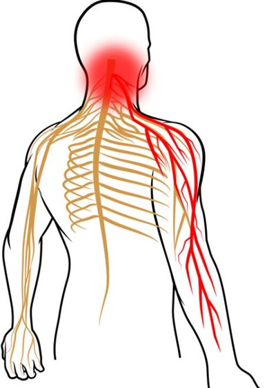
Image on this page provided by ChiroUp.
Coordinated Care, Under One Roof
Chiropractic, physical therapy, acupuncture, and massage therapy are coordinated under one roof with the rest of your treatment plan rather than operating in isolation. Schedule your appointment to get started.