


Disc Herniation, Lumbar Disc Lesion, Stenosis, Pars Fracture, and Arthritis: What Type of Back Pain Do You Have — and What Actually Helps?
By Dr. Matthew Wilson, DC, FIACA | Ashworth Chiropractic, Physical Therapy & Acupuncture | West Des Moines, Iowa
BACK PAIN SERIES — PART 2 OF 4
This is Part 2 of a four-part series we are working on. Each will be linked as they are published online.
Part 1 covers what is the best non-surgical treatment for back pain.
Part 3 covers what you need to think about before you have a back surgery.
Part 4 covers what happens if that surgery to fix your pain.
In Part 1 of this series, we covered the best non-surgical treatments for back pain by stage — acute, subacute, and chronic. But stage is only half the picture. What’s causing your back pain matters just as much as how long you’ve had it.
Disc herniation. Spinal stenosis. Pars fracture. Lumbar osteoarthritis. These are four of the most common specific structural causes of back pain — and they respond quite differently to conservative care. The treatment that works beautifully for one can be wrong for another.
This post goes condition by condition. For each one, we’ll tell you what’s actually happening in the tissue, what conservative care can realistically accomplish, and what the evidence shows. No overselling. No false promises. Just the honest picture.
⚠️ This post is educational and does not replace a clinical evaluation. If you are experiencing progressive leg weakness, loss of reflexes, or any bowel or bladder changes, seek immediate evaluation.
Lumbar Disc Herniation or Lesion — The Good News Most Patients Never Hear
Lumbar disc lesion is another term for the same condition — used clinically to describe disc damage ranging from a minor annular tear through to full herniation
Let’s start with the fact that changes things for most people who hear the words ‘herniated disc’: in the majority of cases, lumbar disc herniations get better without surgery. Not because patients push through the pain and it resolves. But because the disc material actually reabsorbs.
The disc — the cartilage cushion between your vertebrae — is made of a tough outer ring (the annulus fibrosus) and a soft inner core (the nucleus pulposus). When the outer ring tears, that inner material can push out and compress the nearby nerve root. That compression causes the pain, the sciatica, the leg symptoms.
Here’s what most people don’t know: that herniated material is biologically recognized by your body as foreign. Your immune system sends macrophages to clean it up. The disc shrinks. The nerve compression reduces. The symptoms improve.
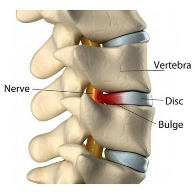
Disc Herniation
What the Research Shows — 66.66% Spontaneous Resorption Rate
Based on articles retrieved from PubMed, a 2017 meta-analysis published in Pain Physician (Zhong et al.) analyzed 11 cohort studies on spontaneous resorption of lumbar disc herniation. The overall incidence of spontaneous resorption with conservative treatment was 66.66% — meaning roughly two out of three disc herniations reabsorb with non-surgical care. In the UK cohort, the resorption rate was 82.94%. Larger herniations — sequestered or extruded discs — tended to reabsorb at higher rates than contained bulges.
That last point is counterintuitive and worth emphasizing: the bigger and more dramatic the herniation on MRI, the more likely it is to reabsorb. The disc material that has fully escaped the annular ring has more contact with the vascular immune system, which accelerates its breakdown.
Zhong M, et al. Incidence of Spontaneous Resorption of Lumbar Disc Herniation: A Meta-Analysis. Pain Physician. 2017;20(1):E45-E52. DOI: https://pubmed.ncbi.nlm.nih.gov/28072796/
The MRI Is Not the Whole Story — And This Is Critical
Here’s something that changes the conversation for a lot of patients: degenerative disc findings on MRI are extremely common in people who have no pain at all. Based on articles retrieved from PubMed, a systematic review of 33 studies covering 3,110 pain-free individuals — published in the American Journal of Neuroradiology from Mayo Clinic’s radiology department (Brinjikji et al., 2015) — found that disc degeneration was present in 37% of asymptomatic 20-year-olds, rising to 96% of asymptomatic 80-year-olds. Disc protrusion was present in 29% of pain-free 20-year-olds. The authors concluded that many imaging-based degenerative features are likely part of normal aging and unassociated with pain.
This matters enormously for how you interpret your imaging — and for decisions about surgery. An MRI is a picture taken at one moment in time, with the spine in an unloaded position lying flat. It shows anatomy. It does not show which structure is generating your pain.
One of the most common scenarios we see in our office is a patient with sciatica-like leg pain, an MRI showing a disc herniation, and a surgical referral — when what’s actually driving the symptoms is hip misalignment, piriformis syndrome, or movement patterns that have progressively tightened the sciatic nerve through the surrounding soft tissue. The disc finding is real. But in many of these cases it’s an incidental finding or a complicating secondary factor — not the primary pain generator. A thorough neurological and orthopedic physical examination is what actually identifies where the pain is coming from. The MRI tells you what’s there. The clinical exam tells you what’s causing the problem.
Patients understandably cling to imaging findings — because an MRI gives a name to the pain, they can see it, and that feels like an answer. We understand that. But the answer to “what is my pain generator” requires a different kind of investigation. Before any surgical decision is made for a disc-related complaint, the structural finding should be correlated with a careful clinical exam that confirms the disc — and not something else — is actually responsible for the symptoms.
Brinjikji W, et al. Systematic literature review of imaging features of spinal degeneration in asymptomatic populations. AJNR Am J Neuroradiol. 2015;36(4):811-816. DOI: https://doi.org/10.3174/ajnr.A4173
What Conservative Care Does for Disc Injuries
The question isn’t whether conservative care works — it often does, and dramatically. The question is whether we can reduce your pain and improve your function while the natural resorption process runs its course.
At Ashworth, disc herniation care is built around several components:
- Directional preference / McKenzie approach — Most disc herniations respond to movement in one specific direction. Identifying that direction and using it consistently reduces nerve compression, centralizes leg pain, and accelerates recovery. This is one of the most important tools in disc rehabilitation.
- Specific spinal manipulation — Not aggressive rotation, but targeted low-force techniques appropriate to the disc presentation. Chiropractic care for disc injuries has a good evidence base and does not worsen herniations when applied correctly. One technique particularly well-suited to disc herniation is lumbar flexion-distraction — a low-force, non-thrust approach used at Ashworth that gently distracts and flexes the lumbar spine while the patient lies prone. Biomechanical research has documented that flexion-distraction reduces intradiscal pressure by up to 65% and enlarges the disc space by 1.5 to 3mm, creating a decompressive effect that reduces nerve root irritation — making it one of the most appropriate manual techniques for acute disc presentations with radiculopathy.
- Postural Restoration (PRI) — Addresses the hip and pelvic asymmetries that place uneven compressive load on the disc. Getting the pelvis into a more balanced position reduces the mechanical stress that keeps the disc aggravated.
- Acupuncture — For pain modulation and reducing the neurological sensitivity (central sensitization) that often accompanies disc-related sciatica.
- Progressive exercise — Building the core stability and movement patterns that protect the disc from re-injury once the acute phase resolves.
💡 The honest timeline: disc-related symptoms often take 6 to 12 weeks to meaningfully improve. Some cases resolve faster. Some take longer. The patients who do worst are those who either do nothing (waiting for spontaneous resorption without managing the nerve irritation) or who pursue surgery before giving conservative care a genuine trial.
When Disc Herniation Actually Needs Surgery
This is important to say directly: some disc herniations do require surgery. The clearest indications are:
- Progressive motor weakness — Foot drop, significant leg weakness that is worsening despite conservative care
- Cauda equina syndrome — Bilateral leg symptoms, bowel or bladder dysfunction, saddle anesthesia. This is a medical emergency requiring immediate surgical evaluation.
- Severe, unrelenting pain that is not responding to any conservative measure after 6 to 12 weeks of appropriate care
For the vast majority of disc herniation patients, surgery is not indicated as a first step — and the evidence supports waiting and treating conservatively first. But when the above criteria are met, we will tell you clearly and refer appropriately.
⚠️ If you have any change in bowel or bladder function alongside back or leg pain, do not wait for a scheduled appointment. Seek emergency evaluation immediately.
Spinal Stenosis — Conservative Care Has More to Offer Than You’ve Been Told
Lumbar spinal stenosis is narrowing of the spinal canal — the space through which the spinal cord and nerve roots travel. As the spine degenerates with age, bone spurs, thickened ligaments, and collapsed disc spaces can all reduce that canal space and compress the neural structures inside.
The classic presentation is neurogenic claudication: leg pain, heaviness, cramping, or weakness that comes on with walking or prolonged standing and is relieved by sitting or bending forward. Many patients describe leaning on a shopping cart as the thing that lets them walk longer — that forward flexion opens the canal and temporarily relieves the compression.
Stenosis is most common in adults over 60, and it’s one of the most frequent reasons for spine surgery in that age group. But the surgery versus conservative care decision is more nuanced than most patients are led to believe.
What the Research Shows on Surgery vs. Conservative Care
Based on articles retrieved from PubMed, a 2016 Cochrane systematic review (Zaina et al.) analyzed five randomized controlled trials covering 643 participants and compared surgical decompression directly against conservative treatment for lumbar spinal stenosis. The findings are more balanced than most surgical referrals suggest:
- At 6 months and 1 year, no significant differences in disability were found between surgical and conservative groups
- At 24 months, surgical decompression showed a modest advantage in disability scores
- Surgical complication rates ranged from 10% to 24%, including fracture, stroke, haematoma, respiratory distress, and reoperation
- Conservative care groups reported zero serious adverse effects across all included trials
The authors concluded they had very little confidence in recommending surgery over conservative care, and specifically noted that clinicians should be careful in informing patients about their options given the complication profile of surgery and the absence of clear benefit in the short and medium term.
Zaina F, et al. Surgical versus non-surgical treatment for lumbar spinal stenosis. Cochrane Database Syst Rev. 2016;1(1):CD010264. DOI: https://doi.org/10.1002/14651858.CD010264.pub2
Conservative Care for Stenosis — What We Do
We want to be upfront about what conservative care for stenosis can and cannot do. It cannot reverse the structural narrowing — the degeneration has occurred and the canal space is what it is. What it can do is manage symptoms, maximize functional walking tolerance, and slow the progression that comes with deconditioning.
The approach at Ashworth:
- Flexion-biased physical therapy — Stenosis patients tolerate flexion better than extension. Kelly Brown Gross, PT, WCS develops programs that exploit this preference to maximize walking tolerance and functional endurance.
- Core stability training — Building the muscular support around the degenerating spine reduces the dynamic compression that occurs with walking and standing.
- Chiropractic mobilization — Gentle, appropriate to the patient’s age and bone quality. The goal is maintaining mobility and reducing the secondary muscle guarding that amplifies stenotic symptoms.
- Acupuncture — Increasingly supported for neurogenic pain modulation in stenosis, particularly for the leg symptoms.
- Activity and lifestyle guidance — Understanding which positions and activities are tolerable versus provocative, and building a sustainable activity plan around that.
Stenosis is often a condition we manage rather than cure. The goal is to keep you as functional and active as possible for as long as possible — and to make sure surgery is genuinely indicated before you pursue it, rather than assumed to be inevitable.
A clinical note on chiropractic technique for stenosis: not all spinal manipulation is appropriate for this condition. High-velocity, low-amplitude (HVLA) thrust techniques — the kind used in Gonstead or Diversified chiropractic — can load the posterior spinal structures in a way that aggravates stenotic symptoms and causes a significant flare. This is one of the most common reasons people report that “chiropractic made it worse.” At Ashworth, we use lumbar flexion-distraction as the primary technique for stenosis, combined with instrument-assisted adjusting of specific segments when indicated and patient-specific exercise.
This approach is consistent with the multimodal care studied by Ammendolia et al. (2015) in the Journal of Manipulative and Physiological Therapeutics, which specifically included lumbar flexion-distraction and produced clinically meaningful improvements in pain, disability, and walking ability in neurogenic claudication patients.
Pars Fracture (Spondylolysis / Spondylolisthesis) — The Back Injury Athletes Often Don’t Know They Have
A pars fracture is a stress fracture in the pars interarticularis — the narrow bridge of bone connecting the front and back elements of a vertebra, most commonly at L5. It’s particularly common in young athletes who perform repetitive extension-loading movements: gymnasts, football linemen, soccer goalkeepers, divers, weightlifters, and cricket fast bowlers.
When both sides fracture (bilateral pars defect), the structural connection between the front and rear elements of the vertebra is lost. This allows the vertebral body to slip forward on the one below — a condition called spondylolisthesis. Grade I and II slips (up to 50% forward displacement) are typically manageable conservatively. Grade III and above — and any case with neurological involvement — require surgical consultation.
Here’s what’s worth knowing: pars fractures are one of the most commonly missed diagnoses in young athletes with back pain. Standard X-rays miss them frequently. A CT scan or SPECT scan is often needed for definitive diagnosis. If you have a young athlete with persistent low back pain that’s worse with extension, hyperextension, and not resolving — this diagnosis deserves investigation.
Conservative Care — This Is the Primary Treatment for Most Cases
Surgery for pars fracture is reserved for high-grade spondylolisthesis, progressive slippage, or neurological compromise — all relatively uncommon. For the vast majority of pars fractures, conservative care is the standard of treatment.
This is an area where the Postural Restoration (PRI) training Dr. Matt completed is particularly relevant. Pars injuries almost universally occur in the context of an anterior pelvic tilt and an extension-biased posture — the spine is already living in the position that loaded the pars. Addressing that underlying positional bias is foundational to treatment.
The conservative approach:
- Activity modification during the active phase — Identifying and reducing the extension loading that stresses the fracture site. For athletes, this means specific activity restriction rather than complete rest.
- Core stabilization — Building the deep stabilizer strength (transversus abdominis, multifidus) that reduces shear stress across the pars. This is not generic core work — it’s specific stabilization training targeted to the L5 level and the pelvic position.
- Postural Restoration — Addressing the anterior pelvic tilt, extension bias and unilateral dominance that is almost universally present. Getting the pelvis into a more neutral position changes the mechanical loading environment around the fracture.
- Gradual return-to-sport — With specific functional criteria rather than time-based return. Going back to extension-loaded sport before the stabilizers are adequate is how athletes re-injure.
💡 Bracing: Rigid bracing is sometimes used for acute active pars fractures in younger athletes to facilitate healing. This is a conversation to have with us at evaluation — whether a brace is indicated depends on the specific presentation, the acuity, and the imaging findings.
A clinical note on technique: HVLA thrust manipulation over an active pars fracture site is contraindicated — it directly loads the posterior structure that is injured. Lumbar flexion-distraction is the more appropriate chiropractic tool for pars presentations: its flexion bias and gentle traction decompress rather than load the pars, and it avoids the rotational forces that aggravate posterior element stress fractures. At Ashworth, flexion-distraction is used as the foundation of manual care for pars fractures, followed by instrument-assisted adjusting of adjacent segments when indicated, and patient-specific stabilization exercise.
Lumbar Osteoarthritis and Facet Joint Pain — Managing Something That Won’t Reverse
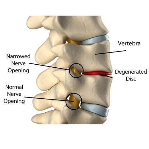
Osteoarthritis in the low back
Lumbar osteoarthritis — degenerative changes in the facet joints (the small joints on the back of the spine) — is one of the most common contributors to chronic low back pain, particularly in adults over 50. The cartilage in these joints thins. Bone spurs form. The joint space narrows. The surrounding soft tissue becomes chronically inflamed and guarded.
The typical presentation is fairly recognizable: stiffness and pain that is worst in the morning, loosens up with movement, worsens again with prolonged standing or extension, and often has a deep aching quality localized to the low back rather than radiating into the leg.
Here’s something worth saying clearly: the relationship between what shows up on imaging and how much pain a patient is in is weak. We regularly see patients with significant facet arthritis on MRI who have minimal symptoms with appropriate management. And we see patients with relatively mild imaging changes who are significantly limited. The image is not the whole story.
What Conservative Care Can Do for Facet Arthritis
Osteoarthritis is a degenerative process. It doesn’t reverse. We’re not going to tell you otherwise. But the pain and functional limitation that come with it are highly manageable — and the evidence consistently shows that active, targeted treatment outperforms passive management and medication alone.
The approach at Ashworth for facet-mediated pain:
- Spinal manipulation and mobilization — Chiropractic care directly addresses the joint restriction and mechanical dysfunction associated with facet arthritis. Restoring motion to restricted segments reduces the load concentration on the most arthritic joints and provides meaningful pain relief.
- Targeted progressive exercise — This is the single most evidence-supported long-term management strategy for lumbar osteoarthritis. Building the muscular support around the arthritic joints reduces the mechanical stress on the degenerated surfaces. Kelly Brown Gross, PT, WCS designs individualized programs around your specific deficits — not a generic handout.
- Graston Technique soft tissue work — The chronic paraspinal muscle tension that accompanies facet arthritis responds well to Graston-based soft tissue mobilization. Dr. Matt holds a Graston M1 certification and uses it specifically for the myofascial component of arthritic back pain.
- Laser therapy — Low-level laser therapy has a growing evidence base for pain modulation in osteoarthritic conditions. It is used at Ashworth as part of a comprehensive care plan when the arthritic pain has an active inflammatory component.
- Lifestyle and load management — Understanding which activities aggravate facet arthritis and which support it, and building a sustainable approach to staying active over the long term.
Facet arthritis is typically a condition we partner with you on for the long term — not fix in a course of care. The goal is maintaining function, managing flares, and preventing the deconditioning spiral that turns manageable arthritis into debilitating limitation.
A word on chiropractic technique for facet arthritis: the same reason HVLA manipulation can flare facet arthritis — the rapid loading of joint surfaces that are already degenerative and inflamed — is exactly why technique selection matters here. Extension-biased thrust techniques often worsen facet pain acutely. Lumbar flexion-distraction is generally better tolerated by facet presentations, and combining it with instrument-assisted adjusting of specific segments produces more consistent relief than traditional thrust manipulation alone. If you’ve had a bad experience with chiropractic for back pain, it is worth asking whether the technique used was appropriate for what was actually wrong with your back.
What If Conservative Care Has Run Its Course?
For most people reading this, conservative care is the right first step — and often the only step needed. But some back pain presentations do eventually lead to surgery. And when that happens, what you do before and after the procedure matters enormously.
Part 3 of this series covers exactly that: ‘You Need Back Surgery — Now What?’ It’s a complete guide to prehabilitation, surgical preparation, and how to give yourself the best possible chance of a good outcome.
And Part 4 addresses one of the most underserved situations in all of musculoskeletal medicine: ‘My Back Still Hurts After Surgery — What Do I Do Now?’ If surgery didn’t provide the relief you expected, that post explains why — and what conservative care can actually accomplish after the operation.
Common Questions
My MRI shows a disc herniation but I’m not sure it’s causing my pain. How do we figure that out?
This is one of the most important questions in spine care. Disc herniations are common incidental findings on MRI — meaning they show up on imaging in people who have no symptoms at all. The key is correlating the imaging findings with the clinical presentation: the location of your pain, whether you have leg symptoms and where they go, what makes it better and worse, and what the neurological exam shows. That correlation is what a clinical evaluation gives you. The MRI alone does not answer the question.
Can a herniated disc fully heal?
In many cases, yes — in the sense that the disc material reabsorbs and symptoms resolve. The Zhong et al. meta-analysis found a 66.66% resorption rate. The disc itself will have some degree of permanent structural change, but that structural change does not necessarily mean ongoing pain. Many people with ‘healed’ disc herniations have residual changes on imaging and no symptoms.
I have stenosis and my surgeon is recommending decompression surgery. Should I get a second opinion?
That is always a reasonable thing to do for elective spine surgery. The Cochrane review we cited found no clear benefit of surgery over conservative care at 6 months and 1 year, with surgical complication rates of 10-24%. A second opinion from both a surgeon and a conservative care provider is appropriate before proceeding with elective decompression.
How do I know if my back pain is from my disc, my facet joints, or something else?
Honestly, distinguishing between these sources clinically is something that requires an evaluation — and even then, there is frequently overlap. Disc pain tends to be worse with flexion and sitting, often refers into the leg. Facet pain tends to be worse with extension and standing, typically stays in the back. Stenosis produces neurogenic claudication that comes on with walking. But these are patterns — not rules — and many patients have multiple contributing sources simultaneously. That’s exactly what a thorough clinical assessment is designed to sort out.
➡️ Next in this series — Part 3: You Need Back Surgery — Now What? A guide to prehabilitation, surgical preparation, and making the procedure actually work.
Clinical References
The following peer-reviewed studies were retrieved from PubMed and cited in this article.
- Zhong M, et al. Incidence of Spontaneous Resorption of Lumbar Disc Herniation: A Meta-Analysis. Pain Physician. 2017;20(1):E45-E52. DOI: https://pubmed.ncbi.nlm.nih.gov/28072796/
- Shen X, et al. Predictive Factors for Resorption in Lumbar Disc Herniation: A Systematic Review. J Inflamm Res. 2026;19:559719. DOI: https://doi.org/10.2147/JIR.S559719
- Zaina F, et al. Surgical versus non-surgical treatment for lumbar spinal stenosis. Cochrane Database Syst Rev. 2016;1(1):CD010264. DOI: https://doi.org/10.1002/14651858.CD010264.pub2
- Gay RE, et al. Intradiscal pressure during flexion-distraction technique: biomechanical research documenting 65% reduction in intradiscal pressure. Referenced in: Park S-H, et al. Effects of the flexion-distraction technique and drop technique on straight leg raising angle and intervertebral disc height of patients with an intervertebral disc herniation. J Phys Ther Sci. 2019;31(8):666-669. PMC: https://pmc.ncbi.nlm.nih.gov/articles/PMC6698474/
- Ammendolia C, Chow N. Clinical outcomes for neurogenic claudication using a multimodal program for lumbar spinal stenosis: a retrospective study. J Manipulative Physiol Ther. 2015;38(3):188-194. DOI: https://doi.org/10.1016/j.jmpt.2014.12.006
About the Author
Dr. Matthew Wilson, DC, FIACA, CCWP is the lead clinician at Ashworth Chiropractic, Physical Therapy & Acupuncture in West Des Moines, Iowa. He is a Palmer College of Chiropractic honors graduate with advanced training in Postural Restoration (PRI), Graston Technique, acupuncture, dry needling, laser therapy, and functional medicine. He works alongside Kelly Brown Gross, PT, MPT, WCS to provide integrated, coordinated care for back pain patients across the full spectrum of presentations and diagnoses.